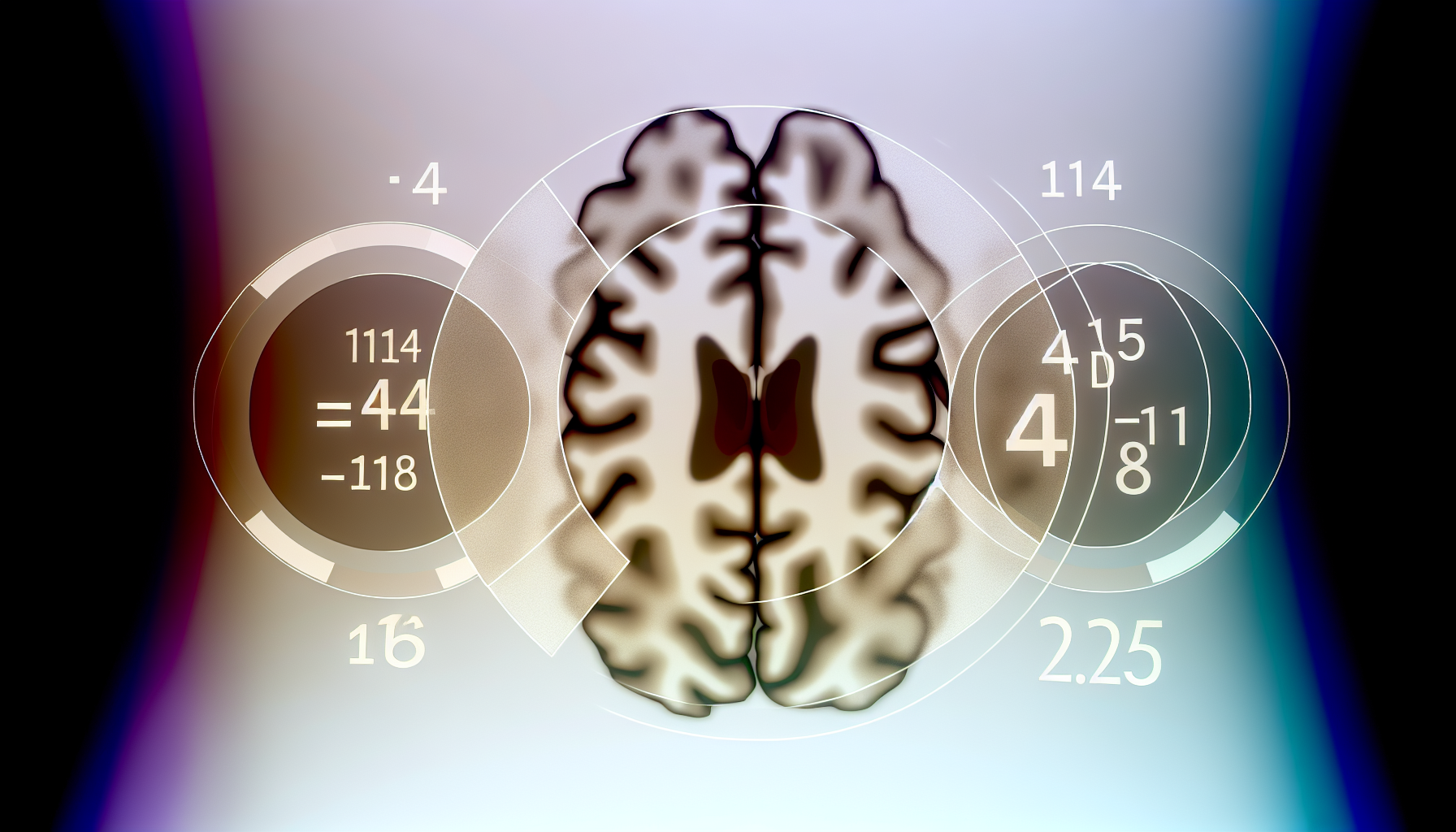
Children’s brains with ADHD do show measurable structural differences in gray matter—once you remove the scanner noise that has muddied results for years. Using a traveling-subject harmonization method on multi-site MRI, researchers uncovered frontotemporal reductions in ADHD, including a robust right middle temporal gyrus signal (beta=-0.255; FDR p=0.001), after correcting measurement bias across scanners [1]. The peer-reviewed study, published August 8, 2025, analyzed scans from 14 “travelers,” 178 typically developing children, and 116 children with ADHD to isolate biological differences from machine effects [1].
Key Takeaways
– Shows TS harmonization exposed ADHD-related frontotemporal gray-matter reductions, with right middle temporal gyrus beta=-0.255 and FDR p=0.001 in children. – Reveals robust multi-site comparability: 14 healthy travelers scanned on four MRI machines over three months removed machine bias while preserving biological sampling. – Demonstrates consistent group-level difference: 116 ADHD versus 178 typically developing children showed smaller frontotemporal gray-matter volumes after TS-corrected analyses. – Indicates TS outperformed ComBat for this goal: TS reduced measurement bias, while ComBat also reduced sampling variation, diminishing ADHD effects across sites. – Suggests clinical potential: TS-enabled gray-matter markers could aid earlier diagnosis and personalized treatment as databases scale toward 1,000 pediatric MRI participants.
How a traveling-subject method cut through MRI noise in ADHD gray matter differences
A key obstacle in multi-site brain imaging is measurement bias—systematic differences introduced by different scanners, sequences, or sites. The traveling-subject (TS) approach answers this by scanning the same healthy individuals across multiple machines, letting researchers estimate and remove machine effects without erasing true biological variance [2]. In this study, 14 healthy “travelers” were scanned across four MRI machines over three months to build the correction model [4]. This design separates measurement bias from sampling differences so disease-related signals can emerge more clearly [2].
Crucially, the team contrasted TS with ComBat, a widely used harmonization technique. While ComBat can reduce site effects, it also dampens sampling variation—the natural differences between groups that researchers actually want to measure—potentially washing out diagnostic signals [2]. In this project, TS preserved biological sampling while removing machine bias, enabling a cleaner comparison of ADHD and typically developing groups across sites [2]. The validation across four scanners confirms the method’s robustness for multi-center pediatric MRI [4].
Quantifying frontotemporal ADHD gray matter changes: the right middle temporal gyrus signal
After TS correction, the clearest ADHD-related gray-matter difference appeared in frontotemporal regions, with the right middle temporal gyrus showing an effect size of beta=-0.255 and an FDR-corrected p=0.001 [1]. This statistic indicates smaller gray-matter volume in that region among children with ADHD compared with typically developing peers, surviving stringent multiple-comparison control across the brain [1]. Independent reporting emphasized that frontotemporal gray-matter decreases were the primary pattern distinguishing the ADHD group when the data were properly harmonized [5].
The sample included 116 children with ADHD and 178 typically developing participants, enabling group-level contrasts that were previously muddied by inter-scanner variability [1]. The right middle temporal gyrus is part of a broader frontotemporal system implicated by the team, suggesting that the differences are not an isolated voxel-level blip but a regionally coherent signal once noise is suppressed [1]. This alignment across analytic pipelines and reports signals a reproducible structural signature under TS harmonization [5].
Why harmonization matters more than bigger samples in ADHD gray matter research
For years, multi-site ADHD MRI studies delivered mixed or null results, often prompting calls for ever-larger cohorts. This work suggests that removing measurement bias may be as important as increasing sample size when effect sizes are modest [1]. By anchoring scanner effects with real people scanned at each site, the TS framework reduces variance where it doesn’t belong—at the machine level—without flattening the meaningful, group-level differences researchers are looking for [2].
The method’s reliability was further shown by consistent performance across four independent scanners during the three-month traveling-subject campaign [4]. Associate Professor Yoshifumi Mizuno described TS as “effective” for harmonizing datasets and enabling reliable identification of ADHD-related frontotemporal volume decreases, contrasting it with ComBat’s tendency to reduce sampling variation [2]. In short, TS improves the signal-to-noise ratio of multi-site analyses without inadvertently silencing the biological signal of interest [2].
ADHD gray matter signals confirmed across datasets and reported by multiple outlets
The study, published August 8, 2025 in Molecular Psychiatry, employed the Child Developmental MRI database, with a long-term goal of reaching 1,000 pediatric participants to strengthen statistical power and subgroup analyses [1]. MedicalXpress’ coverage underscored the database’s scale-up plan and emphasized how TS-corrected analyses revealed smaller frontotemporal volumes in ADHD compared with controls [3]. News-Medical detailed the logistics—14 healthy travelers, four scanners, roughly three months—to extract pure measurement bias from the raw data [4].
NeuroscienceNews highlighted the same right middle temporal gyrus result—beta=-0.255 with FDR p=0.001—as a marquee finding in children, and noted validation on an independent dataset within the Child Developmental MRI framework [5]. Together, these reports converge on a consistent message: when harmonization is done with TS, ADHD gray matter differences become clear, replicable, and regionally specific, especially in the frontotemporal cortex [3][5].
Clinical and research implications of smaller frontotemporal ADHD gray matter volumes
Although structural MRI is not yet a clinical diagnostic for ADHD, the TS-enhanced findings revive the prospect of neuroimaging biomarkers, especially when combined with behavioral and cognitive data. The authors and coverage outlets suggest that gray-matter markers revealed by TS could support earlier diagnosis and more personalized treatment stratification once databases scale and models are prospectively validated [3]. Co-authors emphasized that TS may enable reliable neuroimaging biomarkers by more accurately comparing multi-site data, a prerequisite for clinical translation [5].
For clinicians and trial designers, a reproducible imaging endpoint in frontotemporal regions could help define biologically anchored subgroups, track developmental trajectories, or measure response to interventions. With the database aiming for 1,000 participants, the field can test whether these gray-matter signatures refine prognosis or predict medication and behavioral therapy outcomes across diverse clinical presentations [3]. As larger and more diverse data accrue, TS may become a standard pre-processing step for pediatric multi-site MRI [2][3].
Limitations and what to watch as cohorts approach 1,000 scans
The present analysis focuses on children and hinges on cross-site harmonization, not longitudinal change; thus, it does not quantify developmental trajectories or causal mechanisms. Still, the right middle temporal gyrus effect is statistically robust (beta=-0.255; FDR p=0.001), and the frontotemporal pattern was consistent after correcting for scanner effects [1]. Future steps include expanding the cohort toward 1,000 pediatric participants and validating whether the TS-derived signatures hold across age ranges, comorbidities, and clinical subtypes [3].
Methodologically, TS requires a traveling cohort, which adds logistical complexity but confers a powerful advantage: direct estimation of measurement bias in the same individuals across machines [4]. Press materials and authors argue that this preserves the “biological sampling” necessary to detect genuine diagnostic differences—something ComBat may unintentionally suppress by reducing sampling variation alongside measurement bias [2]. As more centers adopt TS or TS-informed harmonization, expect clearer, more reproducible ADHD gray matter results across sites [2][4].
How this changes the ADHD gray matter conversation
The central takeaway is not that ADHD gray matter differences are enormous—rather, that they are real and quantifiable when machine noise is controlled. With 116 ADHD and 178 typically developing children analyzed under TS, the frontotemporal reductions emerge as a reliable group-level feature, anchored by the right middle temporal gyrus statistic (beta=-0.255; FDR p=0.001) [1]. This reframes past ambiguity as a methodological artifact and points to harmonization as the key to unlocking modest but meaningful brain-structure signals in pediatric ADHD [5].
In sum, the TS approach provides a replicable path to multi-site comparability. It leverages 14 travelers across four scanners to map and remove measurement bias over a three-month window, preserving the between-group differences researchers need to study disease biology [4]. With broader uptake and larger cohorts, TS-corrected MRI could underpin robust biomarkers that complement clinical assessment and guide individualized care in ADHD [3][5].
Sources:
[1] Molecular Psychiatry / PubMed – Brain structure characteristics in children with attention-deficit/hyperactivity disorder elucidated using traveling-subject harmonization: https://pubmed.ncbi.nlm.nih.gov/40781545/
[2] EurekAlert! – Novel accurate approach improves understanding of brain structure in children with ADHD: www.eurekalert.org/news-releases/1097119″ target=”_blank” rel=”nofollow noopener noreferrer”>https://www.eurekalert.org/news-releases/1097119 [3] MedicalXpress – MRI correction method improves understanding of brain structure in children with ADHD: https://medicalxpress.com/news/2025-09-mri-method-brain-children-adhd.html
[4] News-Medical.net – New MRI correction method reveals brain structure differences in children with ADHD: www.news-medical.net/news/20250905/New-MRI-correction-method-reveals-brain-structure-differences-in-children-with-ADHD.aspx” target=”_blank” rel=”nofollow noopener noreferrer”>https://www.news-medical.net/news/20250905/New-MRI-correction-method-reveals-brain-structure-differences-in-children-with-ADHD.aspx [5] Neuroscience News – Brain Structure Differences in Children with ADHD Discovered: https://neurosciencenews.com/brain-structure-adhd-29666/
Image generated by DALL-E 3


Leave a Reply