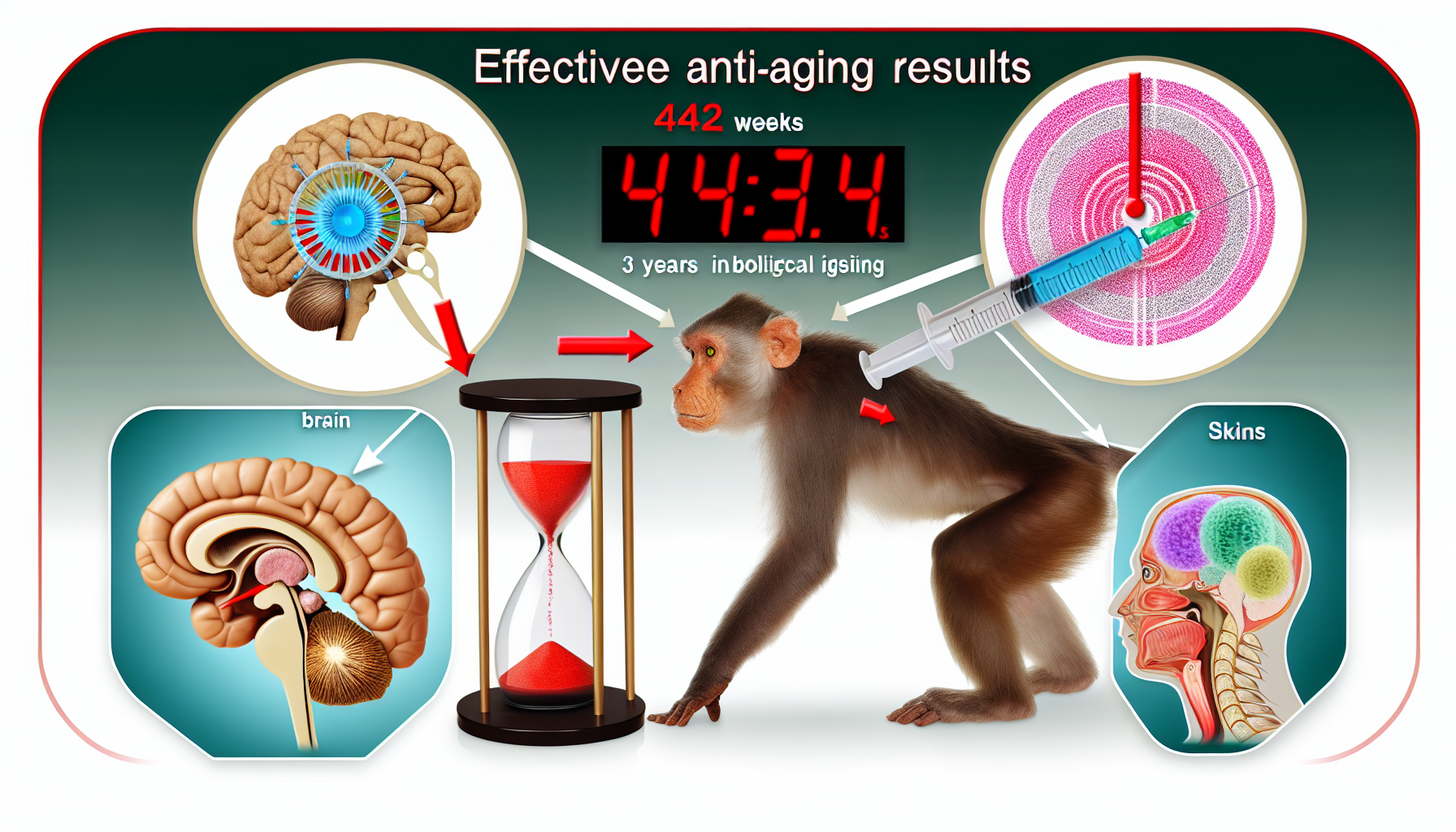
In a 44-week primate study, scientists used senescence-resistant cells—engineered human mesenchymal progenitor cells—to measurably reverse biological aging across multiple organs without detectable safety red flags. Delivered biweekly at 2×10^6 cells/kg, the therapy drove average age reductions of 3.34 years on molecular clocks, alongside cognitive gains, structural brain improvements, and reduced inflammation in aged macaques. The work, published June 13, 2025, in Cell, points to exosome-mediated effects and suggests a potential 5–7-year biological age rollback, while emphasizing the need for careful human translation. [1]
Key Takeaways
– shows average 3.34-year biological age reduction with transcriptomic/DNAm clocks, affecting 54% of tissues after 44 weeks of 2×10^6 cells/kg biweekly infusions. [4] – reveals gene-expression aging clock reversals of 33% in blood, 42% in hippocampus, and 45% in ovary in aged macaques. [2] – demonstrates multi-organ rejuvenation across 61 tissues, including improved cognition, bone density, reduced fibrosis, and restored sperm production without tumorigenicity. [1][3] – indicates decreases in inflammatory SASP markers IL-6 and TNF-α, plus MRI gains in cortical thickness, myelin integrity, and hippocampal connectivity. [5] – suggests exosome-mediated mechanisms and machine-learning clocks estimating 5–7 years of biological age reversal, with no detectable immunogenic responses. [1]
Why senescence-resistant cells target aging at its source
The therapy centers on senescence-resistant cells engineered from human mesenchymal progenitors to withstand stress and suppress the senescence-associated secretory phenotype (SASP), a driver of chronic inflammation and tissue decline. By resisting senescence, these cells release exosomes that modulate inflammatory pathways and rejuvenate neighboring cells’ gene-expression profiles across multiple organs. [1]
Mechanistically, the study points to exosome cargo as a key mediator: small vesicles carrying regulatory RNAs and proteins that dampen IL-6 and TNF-α signaling, helping reset tissue microenvironments from pro-aging to pro-repair states. This correlates with measurable reductions in SASP markers and improvements in structural brain readouts on MRI. [5]
Parallel evidence supports genetic enhancement of stress-resilience pathways. The primate trial included FOXO3-enhanced variants of these mesenchymal progenitors, aligning with known roles of FOXO3 in longevity and cellular repair, and providing a plausible route for sustained anti-senescence activity in vivo. [4]
Inside the 44-week primate trial: dosing, endpoints, and clocks
Aged cynomolgus (crab-eating) macaques received intravenous infusions of senescence-resistant cells biweekly at 2×10^6 cells per kilogram for 44 weeks, enabling longitudinal assessment of safety and efficacy. Investigators profiled 61 tissues to map systemic changes, from blood and brain to ovary and bone, using multimodal endpoints and machine-learning aging clocks. [1]
Clock-based measures showed robust shifts. Transcriptomic and DNA methylation clocks indicated a mean 3.34-year drop in biological age across tissues, with 54% of surveyed tissues registering younger clock values by study end. These molecular findings were anchored to functional measures, including cognition and MRI-based structural brain indices. [4]
Complementing the average effect, tissue-specific reversals were striking: gene-expression aging clocks reversed by 33% in blood, 42% in hippocampus, and 45% in ovary, signaling both systemic and organ-selective rejuvenation patterns that align with observed functional improvements. [2]
What senescence-resistant cells changed in aging primates
Cognitive performance improved in treated macaques, consistent with MRI findings of increased cortical thickness, enhanced hippocampal connectivity, and better myelin integrity—signals typically eroded with age. These structural and functional gains paralleled molecular clock reversals in the hippocampus, a region central to memory. [1][5]
Systemic anti-inflammatory effects were evident. Serum and tissue markers of the SASP, including IL-6 and TNF-α, fell after treatment, aligning with exosome-mediated immunomodulation. This inflammatory reset likely contributed to downstream benefits in tissue repair, fibrosis resolution, and immune rejuvenation reported in the dataset. [5][4]
Reproductive and musculoskeletal features also shifted positively. Investigators observed restored sperm production, improved osteoporosis metrics, and reduced fibrosis—outcomes that underscore multi-organ impact and the potential of senescence-resistant cells to influence stem cell niches and tissue homeostasis beyond the central nervous system. [3]
Safety profile, delivery, and mechanism signals
Across the 44-week dosing schedule, no tumorigenicity emerged on imaging, pathology, or molecular surveillance, addressing a key translational hurdle for cell therapies in aging. Similarly, investigators reported no detectable immunogenic responses to the senescence-resistant cells, an encouraging sign given repeated biweekly infusions. [4][2]
Exosome signatures provide mechanistic coherence. The engineered cells secreted vesicles that curbed inflammatory signaling and altered transcriptomic states in recipient tissues, consistent with widespread clock reversals across 61 tissues and the concentrated improvements in blood, hippocampus, and ovary. These extracellular carriers offer a plausible, non-integrative mechanism with lower oncogenic risk. [1]
Machine-learning clocks suggested a 5–7-year biological age rollback in treated primates, triangulating with the 3.34-year mean reduction across tissues. While different clocks capture overlapping but distinct aging dimensions, convergence across methods strengthens confidence in a genuine rejuvenation signal beyond assay noise. [1][4]
How far from human translation? Timelines, risks, and constraints
Despite compelling primate data, moving senescence-resistant cells into humans requires rigorous, staged trials to confirm dosing, durability, and safety under comorbid conditions common in older patients. Manufacturing scale, release assays for exosome potency, and long-term surveillance for rare adverse events are pivotal next steps. [5]
A central question is generalizability across human tissues and ages. The macaque data show >50% of tissues adopting younger gene-expression profiles and 54% clock responses, but tissue susceptibilities may differ in humans due to lifetime exposures, medications, and immune histories that could blunt or reshape responses. [3][4]
Regulators will probe tumorigenicity risks, biodistribution, and immunogenicity under real-world variability. Parallel development of acellular exosome products could simplify logistics and safety while preserving mechanistic benefits, yet comparative efficacy versus living-cell infusions remains to be established in head-to-head human studies. [1][5]
Measuring age reversal: clocks, functions, and tissues
Quantifying rejuvenation blended molecular and functional endpoints. Transcriptomic and DNA methylation clocks captured a mean reduction of 3.34 years across tissues and signaled responses in 54% of sites examined, including blood and hippocampus, which also exhibited pronounced functional gains. [4][5]
Tissue granularity matters. Blood clock reversal of 33% indicates systemic remodeling, hippocampal 42% aligns with cognitive benefits and MRI connectivity gains, and ovarian 45% suggests reproductive axis responsiveness—collectively reinforcing that senescence-resistant cells can shift both central and peripheral aging trajectories. [2][5]
The breadth of profiling—61 tissues—provides unusual resolution into organ-specific dynamics, strengthening causal inference that the intervention, not chance fluctuations, drove a multi-organ rejuvenation signature. The observed absence of tumorigenicity and immunogenicity under a sustained, biweekly regimen bolsters the therapeutic index. [1][4]
What to watch next in cellular rejuvenation
First-in-human trials will likely prioritize dose-ranging, biomarkers (IL-6, TNF-α, exosome cargo), and early functional endpoints (mobility, cognition), alongside multimodal clocks to compare organ-specific responses. Manufacturing innovations to standardize exosome yield and potency could accelerate development pathways. [5]
Comparative studies—cells versus purified exosomes, or senescence-resistant versus unmodified mesenchymal cells—will clarify mechanism, durability, and scaling economics. Given the primate signals of 5–7-year biological age rollback and multi-organ benefits, carefully designed human studies could determine whether senescence-resistant cells inaugurate a new class of systemic gerotherapeutics. [1]
Sources:
[1] Cell – Senescence-resistant human mesenchymal progenitor cells counter aging in primates: https://linkinghub.elsevier.com/retrieve/pii/S0092867425005719
[2] EurekAlert! – Restoring youth: Scientists use engineered cells to restore vitality in primates: https://www.eurekalert.org/news-releases/1088662 [3] Chinese Academy of Sciences (news) – Scientists Use Engineered Cells to Combat Aging in Primates: https://english.cas.cn/newsroom/research_news/life/202506/t20250620_1045926.shtml
[4] PubMed / NCBI – Reprogramming aging: genetically enhanced mesenchymal progenitor cells show systemic rejuvenation in primates: https://pubmed.ncbi.nlm.nih.gov/40516525/ [5] Cell Regeneration (SpringerOpen) – Attenuation of primate aging via systemic infusion of senescence-resistant mesenchymal progenitor cells: https://cellregeneration.springeropen.com/articles/10.1186/s13619-025-00248-8
Image generated by DALL-E 3







Leave a Reply